Figure 7.
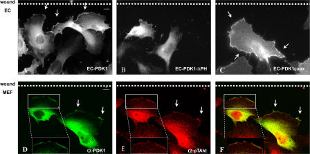
PDK1 localizes at the leading edge of migrating ECs in a PH domain–dependent way and colocalizes with pT308Akt in migrating MEFs. (A–C) ECs infected with indicated retroviruses were seeded at high density on gelatin-coated glass coverslips; monolayer cells were wounded by dragging a plastic pipette tip across the cell surface; and 50 ng/ml VEGF-A was added to the medium. After 6 h, cells were fixed and analyzed by indirect immunofluorescence with mAb α-PDK1 (A and C) or mAb α-myc (B); antigen–antibody complexes were detected with Alexa Fluor 488–conjugated donkey α-mouse IgG. (D–F) MEFs transiently transfected with PDK1 were seeded at high density on gelatin-coated glass coverslips; monolayer cells were wounded by dragging a plastic pipette tip across the cell surface; and medium supplemented with 50 ng/ml PDGF was added. After 6 h, cells were fixed and double stained with mAb α-PDK1 (green) and rabbit α-pT308Akt (red); antigen-antibody complexes were detected with Alexa Fluor 488–conjugated donkey α-mouse IgG and Alexa Fluor 555–conjugated donkey α-rabbit. Images shown are representative of >50% of observed cells. Boxed regions are enlargements of the cell's leading edge. Bars, 10 μm.

