
Figure 6.
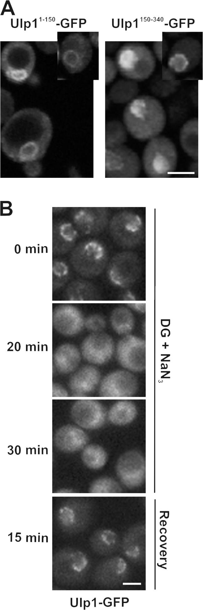
The association of Ulp1p with nuclear pores is energy dependent. (A) The kap binding domains of Ulp1p are targeted to distinct locations. WT cells expressing plasmid-born ULP11-150-GFP or ULP1150-340-GFP were grown to early log phase in selective media, and the distribution of each GFP fusion was examined using confocal microscopy. Individual cells in the culture exhibit different levels of the GFP fusions representative of high and low producers. Insets show cells containing low to moderate levels of the fusion protein. (B) Cells synthesizing Ulp1-GFP (ULP1-GFP integrated at the ULP1 locus) were synchronized in M phase using nocodazole. Cells were washed and placed in media lacking glucose and containing 100 mM 2-deoxyglucose (DG), 10 mM sodium azide (NaN3), and nocodazole. The localization of the GFP fusion was visualized by confocal microscopy after 0, 20, and 30 min of incubation at 30°C. After the 30-min incubation, cells were washed and released into YPD media containing nocodazole (recovery), and the localization of Ulp1-GFP was visualized. Bars, 5 μm.