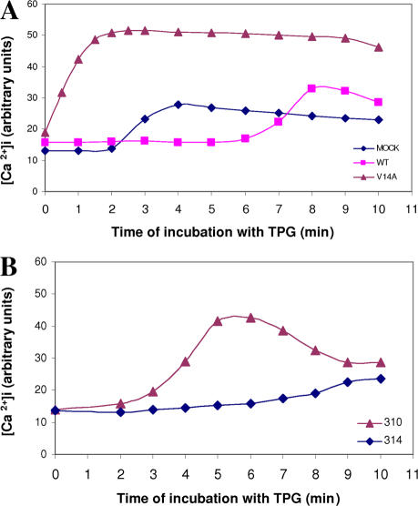
Figure 7.
POSH ubiquitination activity is required for restriction of calcium flow. Quantitative analysis of intracellular calcium before (time 0) and after the addition of 0.5 μM Tpg. (A) Cells transiently transfected with empty (mock), POSH (wild type [WT]), or dominant negative (V14A). (B) H310 and H314 cells. Intracellular free calcium ([Ca2+]i) was detected with the calcium-sensitive dye Fluo-4 and quantified as described in Materials and methods. Each point in the graph represents the Fluo-4 intensity of an image taken at the indicated time point (Fig. S4 shows calcium images of experiment A; available at http://www.jcb.org/cgi/content/full/jcb.200611036/DC1).

