Figure 1.
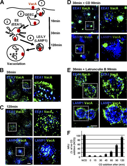
Intracellular trafficking of VacA depends on F-actin. (A) Model for the pinocytosis and intracellular trafficking of VacA as shown previously (Gauthier et al., 2005). (B and C) HeLa cells were intoxicated with VacA at 4°C for 1 h, washed, and incubated for 30 or 120 min at 37°C. Cells were then fixed, permeabilized, and processed for the detection of VacA (green), EEA1, or LAMP1 (blue) by indirect immunofluorescence. Cells were analyzed by confocal microscopy (see Videos 1 and 2 for 3D reconstructions; available at http://www.jcb.org/cgi/content/full/jcb.200609061/DC1). (D and E) Cells were incubated with VacA as in B and C, but, after the 30-min incubation period at 37°C, CD (D) or latrunculin B (E) was added to disrupt the actin cytoskeleton. Cells were further incubated for 90 min before being processed for the detection of VacA (green) and EEA1 or LAMP1 (blue; see Video 3 for 3D reconstructions). (B–E) Images on the right show enlarged views of the boxed regions in the corresponding left panels. All pictures present whole cell reconstructions from confocal sections and represent the merged images of the green (VacA) and blue (indicated endocytic markers) labeling. Bars, 10 μm. (F) HeLa cells were incubated at 4°C with VacA for 1 h and were washed and incubated at 37°C for a total period of 120 min. CD was added immediately or 15, 30, 45, 60, 90, or 120 min after the temperature shift. 5 mM NH4Cl was added to the medium, and cells were incubated for a further 2 h to enable vacuolation to occur. Vacuolation was quantified by neutral red uptake (NRU). Error bars represent SD. NCD, no CD.

