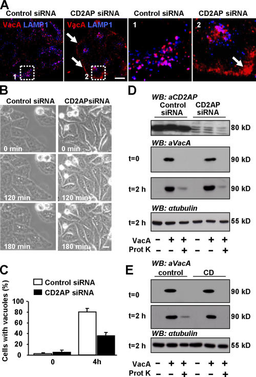
Figure 9.
Down-regulation of CD2AP expression inhibits VacA transfer from GEECs to LEs and VacA-induced vacuolation. (A) 36 h after transfection with control siRNA (left) or CD2AP siRNA (right), HeLa cells were incubated with VacA at 4°C for 1 h, washed, and incubated for 120 min at 37°C. Cells were fixed, permeabilized, and processed for the detection of VacA and LAMP1. Cells were then analyzed by confocal microscopy. Pictures and insets represent the merge between VacA labeling (red) and LAMP1 labeling (blue) from single confocal sections. The boxed areas delineate the regions that are enlarged in the corresponding insets. The arrows show that upon CD2AP depletion, VacA remains in the GEECs. (B) Cells were transfected with control siRNA or CD2AP siRNA. 36 h later, they were incubated at 4°C for 1 h with VacA, washed, and incubated at 37°C for 4 h with DME containing 5 mM NH4Cl. Cells were analyzed by videomicroscopy. (C) Vacuolating cells were scored at the beginning of the film (0) and 4 h later. Results are expressed as the percentage of vacuolating cells among the entire cell population and represent the means ± SEM (error bars) of three independent videos. (D) The amount of intracellular (proteinase K resistant) VacA is identical in cells expressing or not expressing CD2AP. HeLa cells were transfected with control siRNA or CD2AP siRNA as described in Materials and methods. 3 d later, cells were treated with VacA at 4°C for 1 h (t = 0), and internalization of VacA was allowed for 2 h at 37°C (t = 2 h). Cells were then treated with proteinase K as described in Materials and methods to degrade extracellular VacA. The amount of intracellular VacA (i.e., proteinase K resistant) is identical in cells expressing or not expressing CD2AP, indicating that VacA internalization occurs independently of CD2AP. Western blotting against tubulin was used as loading control for the 2-h time point. (E) As a control, cells were pretreated with CD to block VacA pinocytosis as described previously (Gauthier et al., 2005). HeLa cells were pretreated or not pretreated with CD for 20 min before VacA binding and were processed as in D. Bars, 10 μm.

