Figure 2.
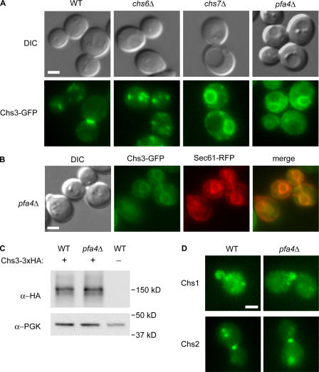
Chs3-GFP mislocalizes to the ER in pfa4Δ cells. (A) Wild-type (KLY9), chs6Δ (KLY1), chs7Δ (KLY3), and pfa4Δ (KLY5) cells expressing Chs3-GFP were observed by differential interference contrast and fluorescence microscopy. (B) Chs3-GFP colocalizes with the ER-marker Sec61-RFP in pfa4Δ (KLY55) cells. (C) Chs3-3xHA levels in wild-type (KLY41) and pfa4Δ (KLY43) cells, as shown by Western blotting with α-HA mAb. α-PGK1 was used as a loading control. (D) Localization of Chs1- and Chs2-GFP in wild-type and pfa4Δ cells. Bars, 2 μm.

