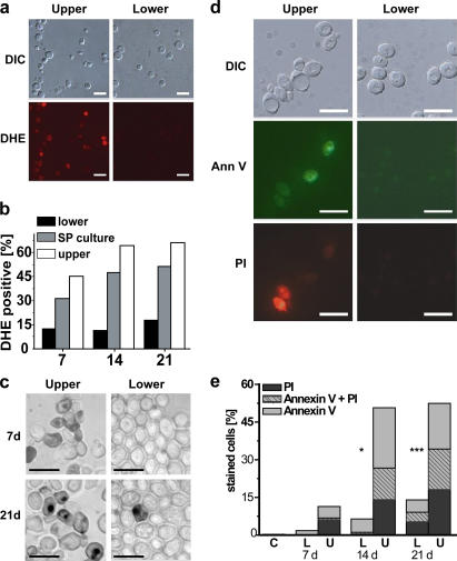
Figure 6.
Evaluation of nonquiescent, upper-fraction and quiescent, lower-fraction cells for ROS, apoptosis, and necrosis. (a) DHE staining to detect ROS in cells from upper and lower fractions from 7-d-old S288c SP cultures. DIC, differential interference contrast. (b) Flow cytometric quantification of DHE-positive, fractionated 7-, 14-, and 21-d-old S288c cells. (c) TUNEL staining to detect DNA fragmentation in 7- and 21-d-old S289 nonquiescent, upper- and quiescent, lower-fraction cells. (d) Ann V and PI costaining of 14-d-old S289 cells. (e) Flow cytometric quantification of 7-, 14-, and 21-d-old S289 lower (L) and upper (U) fraction cells or exponentially growing (C) cells costained with AnnV and PI. Bars, 10 μm.

