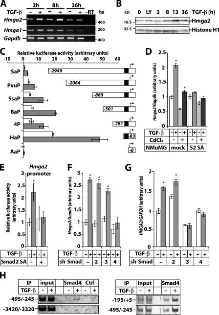
Figure 1.
TGF-β/Smad signaling induces Hmga2 transcription. (A) RT-PCR analysis of Hmga2 and Hmga1 expression in NMuMG cells stimulated with 5 ng/ml TGF-β for the indicated times. (B) Immunoblot analysis of endogenous Hmga2 in NMuMG cells treated with vehicle (0), TGF-β type I receptor inhibitor LY580276 (2.5 μM; LY) for 4 h, or stimulated with TGF-β for the indicated periods of time. Histone H1 serves as a loading control. Molecular mass markers are in bp (A) and kD (B). (C) Hmga2 promoter assays of the indicated deletion constructs in HepG2 cells stimulated (gray bars) or not (white bars) with 5 ng/ml TGF-β for 24 h. The black box in the Hmga2 promoter corresponds to a TCC repeat-rich sequence. (D) Quantitative RT-PCR analysis of Hmga2 expression in NMuMG clones expressing dominant-negative Smad2 (S2 SA) or empty vector (mock) induced or not with 10 μM CdCl2 for 24 h, before stimulation with 5 ng/ml TGF-β for 4 h. (E) Promoter assays of the Hmga2 BaP construct in HepG2 cells transfected with Smad2 SA and stimulated (gray bars) or not (white bars) with 5 ng/ml TGF-β for 24 h. (F and G) Quantitative RT-PCR analysis of Hmga2 expression in NMuMG (F) and MDA-MB-231 (G) clones expressing short hairpin vectors (sh-Smad) directed against Smad2, -3, and -4, or the empty vector and treated with 5 ng/ml TGF-β for 6 h. Asterisks indicate statistically significant gene expression or promoter activity differences between TGF-β–stimulated and nonstimulated conditions (P < 0.05). (H) ChIP assays in NMuMG cells treated or not with 5 ng/ml TGF-β for 2 h using a Smad4 antibody or a preimmune serum (Ctrl) and amplification of Hmga2 promoter fragments.

