Figure 3.
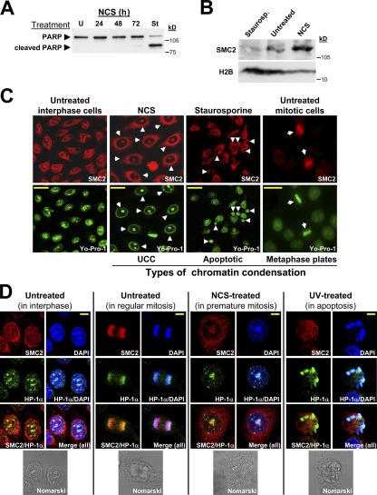
Condensin proteins participate in mitotic chromatin condensation and in UCC but not in apoptotic chromatin condensation. (A) Induction of apoptosis in HeLa cells by staurosporine treatment. PARP-1 cleavage is detected in staurosporine-treated cells (1 μM for 3.5 h). Note the absence of this response in NCS-treated cells (200 ng/ml). U, untreated; St, staurosporine treated. (B) Western blotting analysis of chromatin fractions from NCS- and staurosporine-treated HeLa cells demonstrates condensin recruitment to chromatin in damage-induced UCC but not in apoptotic chromatin condensation. (C) Three types of chromatin condensation in HeLa cells: mitotic, apoptotic, and damage-induced UCC. Immunofluorescence images taken from logarithmically growing HeLa cells treated with 1 μM staurosporine (for 3.5 h) or 200 ng/ml NCS (for 24 h). SMC2, red; DNA (Yo-Pro-1), green. Bars, 20 μm. (D) Visualization of three types of chromatin condensation in HeLa cells stained with both anti-SMC2 (red) and anti–HP-1α (green) antibodies. DNA (DAPI), blue. Bars, 8 μm.

