Figure 1.
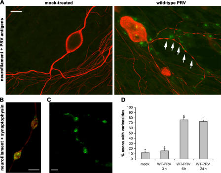
Induction of varicosities along the axons of PRV-infected porcine TG neurons. (A) Confocal images of TG neurons in the inner chamber of mock- or PRV-infected two-chamber models at 24 h after inoculation, stained for the neuronal marker neurofilament 68 (Texas red) and PRV antigens (FITC). Arrows point to varicosities. Bar, 20 μm. (B and C) Confocal images of varicosities in the inner chamber of a two-chamber model at 24 h after inoculation with 2 × 10⁶ PFUs of WT-PRV and double stained for neurofilament (Texas red) and the synaptic vesicle marker synaptophysin (FITC; B) or labeled with FM1-43 (C). Bars, 5 μm. (D) Percentage of axons with varicosities in mock-treated or PRV-inoculated two-chamber models (3, 6, and 24 h after inoculation). Data shown represent means ± SEM of triplicate assays. Percentages indicated by the same letter do not significantly differ (α = 0.05).

