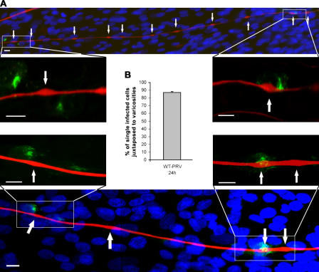
Figure 5.
Varicosities serve as axonal exit sites for PRV. (A) Confocal images of TG neuronal cultures in the inner chamber of PRV-infected two-chamber systems at 24 h after inoculation and stained for neurofilament 68 (Texas red), PRV antigens (FITC), and nuclei (Hoechst 33342). Arrows point to varicosities. Bars, 5 μm. (B) Graph shows the percentage of single-infected cells that are juxtaposed to varicosities, calculated compared with the total number of single-infected cells. Data shown represent means ± SEM of four assays.

