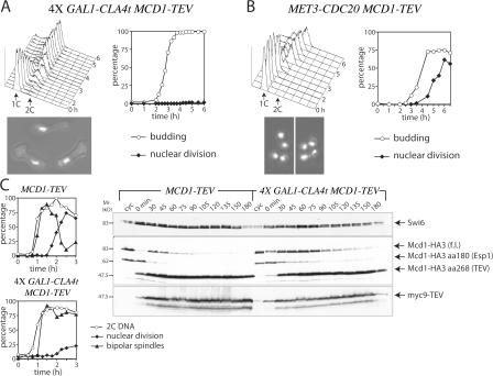
Figure 3.
Mcd1 cleavage is not sufficient for nuclear division upon CLA4t overexpression. (A) GAL1-CLA4t cells expressing Mcd1-TEV and GAL1-TEV (ySP5871) were grown in YEPR at 25°C. Elutriated small G1 cells were released in YEPRG at 25°C (time 0). At the indicated time points, cell samples were analyzed for DNA contents (top left), budding, and nuclear division (top right). Micrographs represent cells at the end of the experiment. (B) MET3-CDC20 MCD1-TEV GAL1-TEV cells (ySP5870) were grown in raffinose medium lacking methionine. Elutriated G1 cells were released in YEPRG containing 2 mM methionine (time 0). Cell samples were analyzed as in A. (C) MCD1-TEV (ySP3448) and GAL1-CLA4t MCD1-TEV (ySP5871) cells were grown in YEPR at 25°C, arrested in G1 by α-factor, and released in YEPRG at 25°C at time 0. Cells were collected at the indicated times for Western blot analysis with anti-HA (Mcd1) and anti-myc (TEV) antibodies (left), FACS analysis of DNA contents (not depicted), and kinetics of nuclear division and bipolar spindle formation (right). Swi6 was used as loading control.

