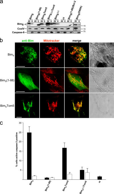
Figure 5.
Subcellular localization of BimS mutants and their pro-apoptotic activity. (a) Localization as determined by cell fractionation. T-REx-293 cells were transiently transfected with wild-type or mutant constructs and induced 7 h later with tet for ∼15 h. Mitochondria (m) and cytosolic fractions (c) were prepared and tested for expression of BimS proteins. As a control (ct) the cells were transfected with the empty expression vector. BimS(1–88)/Bcl-2: cells were cotransfected with BimS(1–88) and an expression construct for hBcl-2. The mutants are described in Fig. 4. (b) Localization of C-terminal mutants as determined by confocal microscopy. Cells were transfected as above, induced 24 h later for 5 h with tet, and BimS was detected by staining with Bim-specific antibodies. Mitochondria were identified by MitoTracker staining. Bars, 10 μm. (c) T-Rex-293 cells were transiently transfected with the constructs indicated (ct, empty vector control). 7 h later, expression of BimS was induced by tet addition as shown. 15 h later, cells were harvested and percentage of active caspase-3–positive cells was determined by flow cytometry. Black bars indicate induction with tet; white bars without tet. The values give the mean of at least three independent experiments/SEM. Expression of transfected proteins was confirmed by Western blotting (not depicted).

