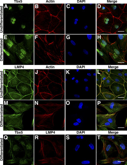
Figure 2.
Localization of Tbx5 and LMP4 during epicardial cell differentiation. HH stage 25 chicken primary epicardial cells were cultured and stained for endogenous Tbx5 (A–H) or LMP4 (I–P) using specific antibodies. (A–D) Undifferentiated epicardial cells stained for Tbx5 (A), Alexa 488 phalloidin pseudocolored red (B), and the nuclear marker DAPI (C). Merged image (D) shows Tbx5 colocalization with DAPI but not with actin-stained phalloidin. (E–H) Epicardial cells induced to differentiate into EPDCs. Tbx5 localization (E) was nuclear but also displayed a filamentous cytoplasmic distribution. Merged image (H) shows Tbx5 colocalization with the nucleus and actin filaments. (I–L) Epicardial cells stained for LMP4 (I), actin (J), and DAPI (K). Merged image (L) shows colocalization of LMP4 with actin. (M–P) Differentiated EPDCs displayed filamentous and cortical localization of LMP4 (M), consistent with the reorganization of actin into stress fibers (N). No significant localization of LMP4 was detected within the nucleus (P). LMP4 also displayed nonfilamentous cytoplasmic localization in undifferentiated and differentiated epicardial cells (I and M). (Q–T) Differentiated EPDCs stained with anti-Tbx5 (Q), anti–LMP4-rhodamine (R), and DAPI (S). Merged image (T) shows significant colocalization of Tbx5 and LMP4 in the cytoplasm along actin filaments. EPDC differentiation was confirmed by the detection of calponin (Fig. S2, available at http://www.jcb.org/cgi/content/full/jcb.200511109/DC1). Bars, 20 μm.

