Figure 3.
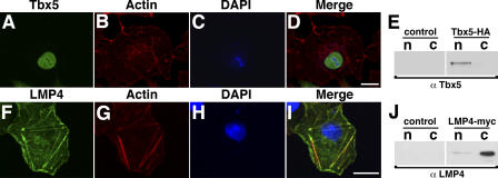
In single transfections, chicken Tbx5 and LMP4 localize to separate cellular compartments. (A–E) COS-7 cells transfected with Tbx5-HA and its expression detected using anti-Tbx5 antibodies (A). Cells were counterstained for actin using Alexa Fluor 633 phalloidin (B) and the nucleus using DAPI (C). The merged image (D) shows Tbx5 exclusively localized to the nucleus. Western blot of fractionated protein lysates from COS-7 cells transfected with Tbx5-HA, revealing exclusive nuclear localization of the Tbx5 protein (E). (F–J) COS-7 cells transfected with LMP4-myc and its expression detected using LMP4 antibodies (F). Cells were counterstained for actin (G) and the nucleus (H) as in B and C, respectively. The merged image (I) shows colocalization of LMP4 to actin stress fibers with no obvious nuclear localization. Western blot of fractionated protein lysates from COS-7 cells transfected with LMP4-myc, indicating localization of LMP4 to the cytoplasm (J). Empty vector transfections were used as controls for cellular fractionation. c, cytoplasmic fraction; n, nuclear fraction. Bars, 20 μm.

