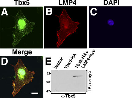
Figure 4.
In cotransfected cells, chicken Tbx5 and LMP4 interact at cytoplasmic sites. (A–D) COS-7 cells cotransfected with Tbx5-HA and LMP4-myc. Cells were stained with anti-HA for Tbx5 (A), anti-myc for LMP4 (B), and the nuclear stain DAPI (C). The merged image (D) shows colocalization of Tbx5 and LMP4 outside the nucleus, predominantly along actin fibers. Coimmunoprecipitation of Tbx5-HA and LMP4-myc from COS-7 protein lysates (E). LMP4-myc was immunoprecipitated with myc antibodies, and the Western blot was processed with Tbx5-specific antibodies. Protein molecular mass markers in kD are indicated on the left of the Western blot. Bar, 20 μm.

