Figure 5.
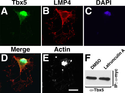
Filamentous actin is required for cytoplasmic Tbx5–LMP4 complex localization. (A–E) COS-7 cells cotransfected with Tbx5-HA and LMP4-myc. 24 h after transfection, cells were treated with 2 μM latrunculin A for 60 min to sequester actin monomers. Cells were processed with anti-HA for Tbx5 (A), anti-myc for LMP4 (B), the nuclear stain DAPI (C), and Alexa Fluor 633 phalloidin to detect actin (E). The merged image (D) shows that the Tbx5–LMP4 complex no longer displays a filamentous pattern. For comparison, actin distribution of the cell in A–D is shown (E). (F) Coimmunoprecipitation of Tbx5 and LMP4 after actin disruption. Tbx5-HA was coprecipitated along with LMP4-myc in lysates from latrunculin A and DMSO control-treated COS-7 cells. Bar, 20 μm.

