Figure 6.
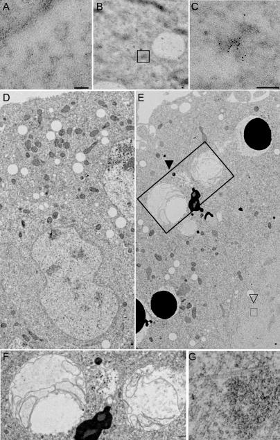
Ultrastructural analysis of Drosophila GWBs and the effect of GW loss on embryos. (A) Thin sections of embryos do not show appreciable immunogold localization when preimmune serum is used. (B and C) Sections stained with α-GW antibodies show appreciable immunogold signal in irregular, electron-dense structures. These are not membrane bound or associated with any other known cytoplasmic structure. Boxed area in B represents a single structure; a representative example is shown at higher magnification in C. (D) Thin sections of wild-type 3-h embryos show characteristic structures (including nuclei) surrounded by a distinct bilayer membrane, which is continuous with the rough endoplasmic reticulum, as well as mitochondria. (E) Homozygous gw 1 3-h mutant embryos have few recognizable nuclei and darkly staining membrane-bound vesicles, presumably corresponding to yolk particles in the embryo cortex, from which they are usually excluded at this later stage of development. Large multivesicular bodies (closed arrowhead and large box) are seen and are shown in higher magnification in F. (G) A higher magnification of the aggregates of filamentous structures indicated by the open arrowhead and small box in E. Bars, 0.2 μm.

