Figure 3.
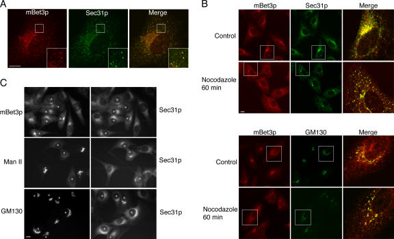
The localization of mBet3p is disrupted by reagents that are known to disrupt the tER. (A) The localization of mBet3p (left, red) largely overlaps (Merge) with Sec31p (middle, green) in BSC-1 cells. The insets are magnified on the bottom right. (B) BSC-1 cells, treated for 1 h with 10 μg/ml nocodazole, were fixed and stained with antibodies directed against mBet3p (red) and Sec31p (top, green), and mBet3p (red) and GM130 (bottom, green). The insets are magnified on the right. (C) The localization of mBet3p clusters in response to microinjected Sar1p H79G, which is a GTP-locked mutant. NRK cells were microinjected with 1.5 mg/ml Sar1p H79G. mBet3p (top), Man II (middle), and GM130 (bottom) were visualized by indirect immunofluorescence. All injected cells are marked with an asterisk. Bars, 10 μm.

