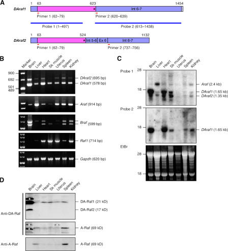
Figure 3.
Expression of DAraf1/2 mRNAs and the proteins in mouse tissues. (A) DAraf1/2 mRNAs together with primers and probes used for RT-PCR and Northern blotting, respectively. Pink boxes, coding regions including termination codons; purple boxes, untranslated regions; Ex, exon; Int, intron. Asterisks represent termination codons. The numbers indicate nucleotide numbers starting from the 5′ ends of the cDNAs. (B) Quantitative RT-PCR detecting DAraf1/2, Araf, Braf, and Raf1 mRNAs. Glyceraldehyde 3-phosphate dehydrogenase (Gapdh) mRNA is shown as a standard. (C) Northern blotting detecting DAraf1/2 and Araf mRNAs with probe 1 and DAraf1 mRNA with probe 2. Ethidium bromide (EtBr) staining of a gel shows 28S and 18S rRNAs as a standard. (D) Immunoblotting detecting DA-Raf1/2 and A-Raf proteins with the anti–DA-Raf pAb. The A-Raf bands were verified by immunoblotting with the anti–A-Raf pAb. Each lane contains 50 μg of proteins. The asterisks indicate unidentified extra protein bands reacted with the anti–DA-Raf pAb.

