Figure 6.
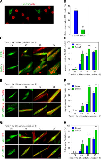
Induction of the myogenic differentiation phenotype in C2C12 cells by highly expressed DA-Raf1. (A) Induction of the cell cycle arrest in C2C12 cells exogenously expressing DAraf1. C2C12 cells were transfected with HA–DAraf1 and incubated with BrdU for 2 h. The anti-HA staining detecting the expression of HA–DA-Raf1 (green) is merged with anti-BrdU staining (red). (B) Ratio of the BrdU-incorporating C2C12 cells in the analysis of A. (C) Facilitation of myogenin expression in C2C12 cells exogenously expressing DAraf1. The cells were cultured in the differentiation medium. Control EGFP fluorescence (green) or anti-HA staining detecting the expression of HA–DA-Raf1 (green) is merged with antimyogenin staining (red). (D) Ratio of the myogenin-expressing cells in the analysis of C. (E) Promotion of MyHC expression in C2C12 cells exogenously expressing DAraf1. Control EGFP fluorescence (green) or anti-HA staining detecting the expression of HA–DA-Raf1 (green) is merged with anti-MyHC staining (red). (F) Ratio of the MyHC-expressing cells in the analysis of E. (G) Facilitation of TnT expression in C2C12 cells exogenously expressing DAraf1. Control EGFP fluorescence (green) or anti-HA staining detecting the expression of HA–DA-Raf1 (green) is merged with anti-TnT staining (red). (H) Ratio of the TnT-expressing cells in the analysis of G. The values in the graphs are means ± SD (error bars) of three independent experiments. *, P < 0.05; **, P < 0.01 by t test compared with the control at each time point. Bars, 20 μm.

