Figure 8.
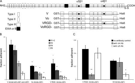
Role of integrins in the adhesion of erythroid progenitors. (A) Schematic diagram of recombinant fibronectin fusion proteins. The V fragment contains both the α4β1- and α5β1-binding sites, Vo contains only the α5β1-binding site, VRGD− contains only the α4β1-binding site, and VoRGD− contains neither integrin-binding site. (B) Characterization of the adhesion of sorted R1 + R2, R3, R4, and R5 cells to 10 μg/ml V, Vo, or VRGD substrates at an acceleration of ∼1,000 g. (C) α4β1 and α5β1 integrins mediate adhesion to different fibronectin domains. Adhesion of sorted R1 + R2 cells to 10 μg/ml recombinant fibronectin fragments Vo or VRGD− with and without pretreatment with function-blocking antibodies to α4 and α5 integrins. Each bar represents the mean fraction of cells adherent in each of three replicate wells. In all cases, error bars are the SD. *, P ≤ 0.05; **, P ≤ 0.01.

