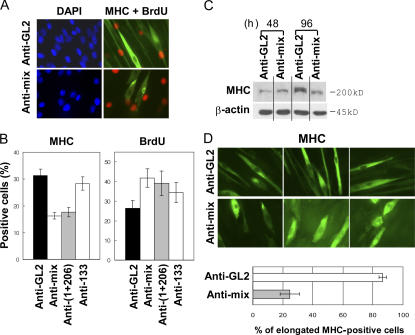
Figure 3.
Inhibition of the miRNAs by 2′-O-methyl antisense oligonucleotides inhibits differentiation. (A) C2C12 cells transfected with antisense oligonucleotide against GL2 or a mixture of antisense oligonucleotides against miR-1, -133, and -206 (anti-mix). Three transfections at 24-h intervals in GM (serum+) were followed by serum depletion (DM). 96 h after serum depletion, MHC expression (green) and BrdU incorporation (red) were detected by immunostaining. Blue indicates nuclei stained by DAPI. (B) Quantification of percentage of BrdU- and MHC-positive cells in samples prepared as described in panel A. Anti-(1+206) indicates a mixture of 2′-O-methyl antisense oligonucleotides against miR-1 and -206. Each value is a mean of triplicate. (C) Immunoblots at 48 and 96 h after serum depletion (DM). The transfection and induction of differentiation were performed as described in A. (D) Cell morphology visualized by MHC immunostaining as described in A (top). Results are quantitated at the bottom. Cells that were elongated were twofold longer than proliferating C2C12. Each value is a mean of triplicate experiments.

