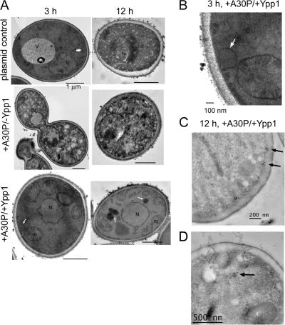
Figure 5.
Transmission electron microscopy images of cells expressing Ypp1p and A30P. (A) S288c cells transformed with pTF302 (A30P) or pTF300 (empty vector) and pTF602 (YPP1) or pTF604 (empty vector) were pregrown in noninducing media to mid-log phase, transferred to inducing media and induced for 3 or 12 h. Top: −A30P/−Ypp1 (cells contained two plasmids with no inserts). Middle: +A30P/−Ypp1 (cells contained two plasmids, one with no insert). Bottom: +A30P/+Ypp1 (cells contained two plasmids). Bars, 1 μm. (B) Magnification of +A30P/+Ypp1 cell at 3 h shows vesicle budding from the plasma membrane. (C and D) Immunogold labeling of cells expressing A30P and overexpressing Ypp1p at 12 h. A secondary antibody conjugated with 50-nm gold particles was used to detect A30P. Several clusters of gold particles occurred in the vicinity of the plasma membrane (C) and also in the interior of cells (D).

