Figure 8.
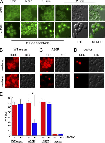
Pheromone protects cells from A30P toxicity. (A) Effect of α-factor on cells expressing Ypp1p-GFP. Top (−α-factor): no change in the localization of Ypp1p-GFP occurred over 20 min. Bottom (+α-factor): 10 μM α-factor was added to cells and then images were acquired from 2 to 20 min. Strain: YPP1-GFP cultured in YPD. Identical instrumental conditions were used for all images. Bar, 5 μm. The ROS assay using the DHR 123 dye is shown in (B) WT α-syn, (C) A30P, and (D) vector control. The FY23 strain was transformed with pTF201 (WT α-syn), pTF202 (A30P), pTF203 (A53T), or pTF200 (empty vector) and pregrown in noninducing media to mid-log phase. 10 μM pheromone was added after shifting to inducing media; after 2 h in inducing media the DHR 123 dye was added to 5 μg/ml; and, before acquiring images at 3 h, cells were washed and resuspended in inducing media. The two images labeled “MERGE” (in A) had brightness and contrast adjustments of +10 and +40, respectively. (E) Plot of mean ROS (%) (± SD). Cells (n = 292–696) were counted in two independent experiments. *, P = 1.1 × 10⁻⁷. DIC, differential interference contrast; DHR, rhodamine 123.

