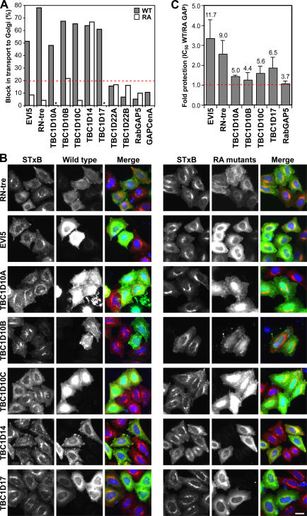
Figure 4.
A subset of human Rab GAPs influence Shiga toxin transport. (A) Shiga toxin uptake assays were performed on HeLa cells transfected with a library of wild-type human Rab GAPs. Those Rab GAPs showing a block in Shiga toxin uptake were retested as catalytically inactive RA mutants. To measure the extent of Shiga toxin uptake, HeLa cells were transfected with the GFP-tagged wild-type and catalytically inactive mutant GAPs indicated in the figure. These cells were then fixed and stained for a Golgi marker. The percentage of untransfected and transfected cells in which STxB had reached the Golgi was counted for each condition, and the specific reduction in Shiga toxin transport was calculated by subtracting these two figures (transfected − untransfected cells). These numbers are plotted on the bar graph. The dotted red line indicates the cut-off chosen for consideration as a positive. Asterisks indicate that transport was the same as in control cells. (B) Images of the Rab GAPs showing a catalytic activity–dependent block or reduction in Shiga toxin uptake after 60 min. Shiga toxin is in red, transfected Rab GAPs are in green, and DNA is stained blue. (C) Cytotoxicity assays were performed using the Rab GAPs listed in the figure. The IC50 of Shiga-like toxin 1 was measured in cells expressing either the wild type (WT) or the catalytically inactive (RA) mutant GAPs. These values were then corrected for transfection efficiency averaging 13.9–18.7%. An increase in the IC50 for wild type compared with RA GAPs indicates reduced cytotoxicity, and this is plotted in the graph as the fold protection, with 1 (red dotted line) corresponding to no protection (n ≥ 3). The corrected IC50 values for each condition are shown above each column, and the IC50 of empty vector–transfected control cells was 3.5 ng/ml. Error bars represent SD. Bar, 10 μm.

