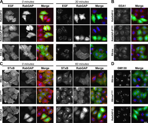
Figure 6.
Discrete Rab GAPs define the Shiga toxin and EGF uptake pathways. (A and B) HeLa cells were transfected for 24 h with RabGAP-5, the catalytically inactive RabGAP-5R165A mutant, or RN-tre. (A) EGF uptake assays were performed, and the initially bound EGF at 0 min of transport and extent of EGF transport at 30 min are shown in the figure. EGF is in red, and transfected Rab GAPs are in green. (B) The cells were fixed and stained for the transfected Rab GAPs (green) and EEA1 (red). (C and D) HeLa cells were transfected for 24 h with RN-tre, the catalytically inactive RN-treR150A mutant, or RabGAP-5. (C) Shiga toxin uptake assays were performed, and the initially bound Shiga toxin at 0 min of transport and extent of STxB transport to the Golgi at 60 min are shown. STxB is in red, and transfected Rab GAPs are in green. (D) The cells were fixed and stained for the transfected Rab GAPs (green) and GM130 (red). DNA is stained blue in all panels. Bar, 10 μm.

