Figure 4.
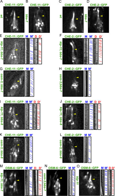
Transport assays of IFT particle subcomplexes A and B in bbs-7 single and bbs-7 or bbs-8; kinesin-2 double mutants. Micrographs of the distribution of IFT-A (CHE-11∷GFP) and -B (CHE-2∷GFP and OSM-6∷GFP) subcomplexes along sensory cilia (CHE-11∷GFP in A, B, E, G, I, and K; CHE-2∷GFP in C, D, F, H, J, and L; and OSM-6∷GFP in M–O). Kymographs and corresponding graphs in E–O (right) show the diagonal lines that represent trajectories of movement along the initial (M and M′) and distal segments (D and D′). Arrowheads point to initial-distal segment junctions. In wild-type (wt) animals (A and C), CHE-11∷GFP and CHE-2∷GFP move identically along initial and distal segments. In bbs-7 mutants (B and D), IFT-A and -B dissociate, CHE-11∷GFP only moves within the initial segment, and CHE-2∷GFP moves along both the initial and distal segments. In klp-11;bbs-7 or bbs-8 double mutants, CHE-11∷GFP, CHE-2∷GFP, and OSM-6∷GFP move at OSM-3–kinesin's fast velocity along the initial and distal segment (E, F, I, J, and M). In osm-3; bbs-7 or bbs-8 double mutants, CHE-11∷GFP, CHE-2∷GFP, and OSM-6∷GFP move at kinesin-II's slow rate in the remaining initial segment (G, H, K, L, and N). OSM-6∷GFP moves at OSM-3's fast rate in klp-11 mutants (O). Unlike in bbs single mutants, IFT particles are stable and do not dissociate into IFT-A and -B in bbs-7 or bbs-8; kinesin-2 double mutants.

