Figure 3.
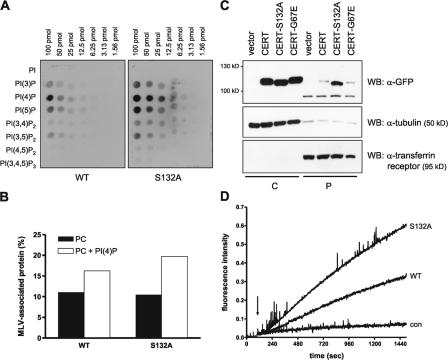
CERT phosphorylation on serine 132 modulates PI(4)P binding and ceramide transfer activity. HEK293T cells transiently expressing the indicated GFP-tagged CERT variants were harvested by hypotonic lysis, and the cytosol fraction was recovered after 100,000 g centrifugation. Samples containing equal amounts of GFP fluorescence were used for protein–lipid overlay (A), flotation (B), and in vitro ceramide transfer assays (D). (A) Phosphatidylinositol phosphate arrays were incubated with cytosol from cells transiently expressing GFP-tagged CERT-WT and -S132A, and bound proteins were detected with GFP-specific primary followed by HRP-labeled secondary antibody. (B) MLVs consisting of PC or PC + 5% PI(4)P were incubated with cytosol and separated by centrifugation. The amount of CERT protein in the top (MLV) and bottom fractions was quantified by measuring GFP fluorescence and set as 100%. Results are plotted as percentages of protein recovered in the MLV fraction. (C) Cytosol (C) and the 100,000 g pellet (P) containing cellular membranes were analyzed by immunoblotting with GFP-specific antibody. The purity of the individual fractions was confirmed by detection of the transferrin receptor in the membrane and tubulin in the cytosolic fraction. (D) Donor liposomes containing TNP-PE and pyrene-ceramide were mixed with unlabeled acceptor liposomes. After 60 s, cytosol from cells transiently expressing GFP-tagged CERT-WT, -S132A, or GFP alone (con) was added, and pyrene fluorescence at 395 nm was recorded.

