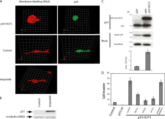
Figure 5.
A375 melanoma cell invasiveness is regulated by p53 activity. (A) Confocal images of A375P cells expressing plasmid EGFP alone or p53 H273. Cells were plated in Matrigel, and a gradient of serum growth factors was established to observe the mode of migration. The transfected cells are visualized using GFP, and plasma membranes were labeled with fluorescent WGA (wheat germ agglutinin; red). (B) p21 induction in A375P cells. A375P cells were untreated or treated with etoposide. Total protein lysates were analyzed by SDS-PAGE followed by immunoblot analysis using antibodies to p21. Normalization was performed with an anti–α-tubulin antibody. (C) RhoA activity in transfected A375P. Cells expressing GFP-tagged p53 H273 or plasmid EGFP (control) were compared for GTP-RhoA levels as in Fig. 2. The image is representative of three independent experiments. The values are the mean ± SD (error bars). (D) Invasiveness in 3D matrigel matrix of A375P-expressing p53 wt or p53 H273 treated with TAT-C3, Y27632, or H1152 as indicated. Results are expressed as in Fig. 4.

