Figure 5.
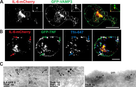
IL-6 and TNFα colocalize in the recycling endosome. (A and B) RAW264.7 cells were transfected with IL-6–mCherry and either GFP-VAMP3 (A) or GFP-TNFα (B) and activated with LPS and IFN for 4 h before fixation. In addition, to label transfected cells with Tfn (blue), macrophages were incubated in the presence of AlexaFluor647-conjugated Tfn (B). Cells were imaged via high resolution confocal microscopy. (A) A large proportion of IL-6–positive structures colocalize with the recycling endosome markers VAMP3 (A), endocytosed Tfn, and TNFα (B). In A, the insets highlight single ring-shaped VAMP3-positive structures containing IL-6. In B, colored arrows are used to highlight three recycling endosome structures containing all three cargo proteins. (C) Immuno-EM of IL-6–GFP, endogenous TfnR, and TNFα labeled with 5, 10, or 15 nm gold particles. Peri-Golgi (left) or peripheral (middle and right) recycling endosomes (RE) are labeled with TfnR, which is recycling from the plasma membrane (pm; right). IL-6–GFP or TNFα (arrows) colocalized with TfnR (arrowheads) in recycling endosomes. G, Golgi complex. Bars (A), 10 μm; (B) 5 μm; (C) 100 nm.

