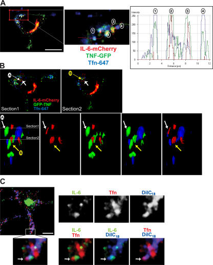
Figure 6.
Compartmentalization of endosomal structures containing IL-6, TNFα, and Tfn. (A–C) Macrophages were transfected with fluorescently tagged IL-6 (C) or both IL-6–mCherry and TNF-GFP (A and B) and were subsequently incubated in the presence of fluorescently conjugated Tfn. In A, a fluorescence intensity line scan profile was generated along the red line, with fluorescence peaks for the four recycling endosomes highlighted. In IL-6–positive structures, the peak fluorescence appears to overlap with the peak of TNFα (structures 2 and 3), but the Tfn fluorescence is more dispersed in these structures compared with those containing TNFα and Tfn alone (structures 1 and 4). (B) Two confocal sections are shown highlighting recycling endosome structures, which are labeled A and B. The enlarged area represents a surface-rendered 3D image of the region highlighted. Images are viewed in the direction of the large arrows and in the cross section through the cell. This demonstrates examples of recycling endosomes with both overlapping and discrete domains for each of the markers. In C, transfected macrophages were additionally subjected to labeling using the DilC18(5)-DS lipophilic dye (blue) before incubation with Tfn (red) as described in Materials and methods. The enlarged area shows Tfn and IL-6 labeling separate domains within a single dye-positive endosomal structure (highlighted by white arrows). Bars (A), 5 μm; (B and C) 10 μm.

