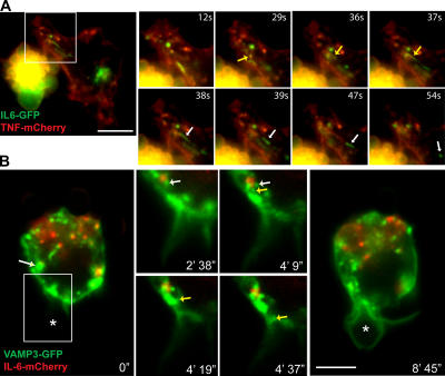
Figure 9.
Trafficking of IL-6 from recycling endosomes can be regulated independently of other recycling endosome functions. (A) Dual-color live imaging of macrophages transfected with IL-6–GFP (green) and TNFα-mCherry (red). Enlarged images demonstrate the tubulation of a structure containing both TNFα and IL-6 (yellow arrows), with IL-6 alone budding off (white arrows) to form a new vesicle. (B) Dual-color live imaging of the phagocytosis of IgG-opsonized 3-μm latex beads by macrophages transfected with VAMP3-GFP (green) and IL-6–mCherry (red). Frames from the dual-color video were extracted to highlight the movement of vesicles containing VAMP3 (yellow arrow) but not IL-6, budding off a recycling endosome containing both cargoes (white arrows), and trafficking toward the phagocytic cup. The phagocytosing bead is highlighted with asterisks. Boxed areas are magnified at the right. Bars (A), 10 μm; (B) 5 μm.

