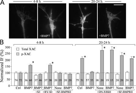
Figure 5.
LIMK and SSH control the phosphorylation of ADF/cofilin in response to BMP7. (A) Representative fluorescent images of Xenopus growth cones of 4–8-h and overnight cultures that were immunostained with a specific antibody against p-XAC without and with bath BMP7 treatment (5 nM for 10 min). (B) Normalized levels of p-XAC and XAC in Xenopus growth cones of 4–8-h cultures and overnight cultures with and without bath BMP7 exposure (5 nM for 10 min) under different treatments. The numbers of growth cones examined for each condition are indicated on the bars. Asterisks indicate values that were significantly different from the control (*, P < 0.01; t test). Error bars represent SEM. Bar, 10 μm.

