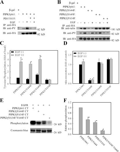
Figure 5.
PIPKIγ661 is tyrosine phosphorylated by EGF stimulation. (A) HeLa cells were transfected with β-gal or HA-PIPKIγ661 separately, pretreated with or without EGFR-specific inhibitor PD153035, and stimulated with 10−9 M EGF for 5 min. Cells were used in immunoprecipitation with anti-HA antibody, and the tyrosine phosphorylation was detected by anti-phosphotyrosine antibody. (B) HA-tagged PIPKIγ661, PIPKIγY644F, PIPKIγY649F, or PIPKIγY634F were transfected into HeLa cells separately and stimulated with 10−9 M EGF for 5 min. Cells were subjected into immunoprecipitation with anti-HA antibody, and the tyrosine phosphorylation was detected. The amount of talin in the immunoprecipitation complex was detected by anti-talin antibody. (C) Tyrosine phosphorylation of HA-tagged PIPKIγ661, PIPKIγY644F, PIPKIγY649F, or PIPKIγY634F with or without EGF stimulation was quantified. (D) Talin association with HA-tagged PIPKIγ661, PIPKIγY644F, PIPKIγY649F, or PIPKIγY634F with or without EGF stimulation was quantified. (E) Reconstituted c-tail of wild-type PIPKIγ661, PIPKIγY644F, PIPKIγY634F, or PIPKIγY634F/Y644F was used as substrate of purified EGFR in the in vitro kinase assay. (F) The relative phosphorylation of wild-type PIPKIγ661, PIPKIγY644F, PIPKIγY634F, or PIPKIγY634F/Y644F was quantified. The phosphorylation of wild-type PIPKIγ661 was set as 100% (*, P < 0.01 compared with wild-type PIPKIγ661). Quantifications are expressed as means ± SEM of three separate experiments.

