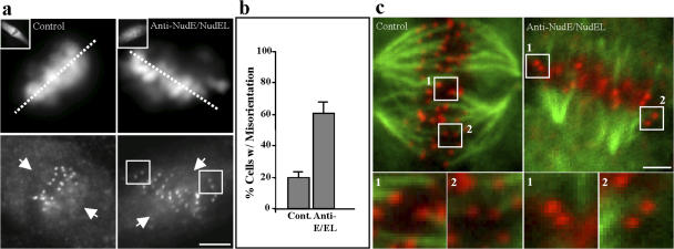
Figure 6.
Effects of anti-NudE/NudEL antibody microinjection on kinetochore orientation and kinetochore microtubule attachment. (a) Mitotic LLC-PK1 cells were injected with anti-NudE/NudEL antibody at nuclear envelope breakdown and allowed to progress to metaphase. Cells were fixed 1 h after injection and processed for immunofluorescence microscopy. Kinetochore pairs (boxed) showed evidence of misorientation relative to the metaphase plate (dotted lines; positions of the spindle poles are indicated by arrows). Insets show staining for the injected anti-NudE/NudEL antibody as a marker for injection. (b) Fraction of cells showing one or more misoriented kinetochore pairs. Error bars represent the SEM. (c) The stability of microtubule attachment at the kinetochore was examined after a 10-min exposure of anti-NudE/NudEL–injected cells to decreased temperatures (0°C). Although microtubule bundles could still be observed, attachment to individual kinetochores was clearly eliminated in some cases, as shown in boxed areas; these are numbered and shown magnified in the insets. Images of control cells in panels a and c were treated with MG132 to prevent anaphase onset. Bars (a), 5 μM; (c) 3 μM.

