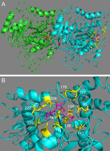
Figure 1.
Location of altered residues. (A) The structures of yeast Tub1 and Tub2 are homology models based on the structure of the αβ-tubulin heterodimer from bovine brain obtained by electron crystallography (Nogales et al., 1998; Richards et al., 2000). The C termini of Tub1 (residues 442–447) and Tub2 (residues 428–457) were not included in the model because they are not resolved in the bovine structure. Tub1, green; Tub2, cyan; GTP bound to Tub1, orange; GDP bound to Tub2, magenta; Tub2 residues changed to alanine in this study, yellow; Tub2-C354, red. (B) The GTP-binding pocket of Tub2. Coloring as in A. Numbers indicate positions of the mutated residues.

