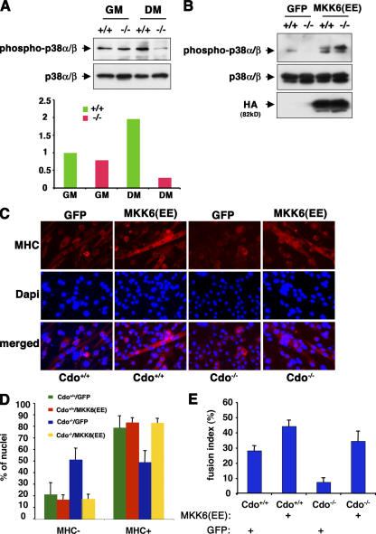
Figure 3.
MKK6EE rescues the differentiation-defective phenotype of Cdo−/− satellite cells. (A, top) Lysates from satellite cells of the indicated Cdo genotype cultured in GM or DM were Western blotted as indicated. (bottom) The image was quantified by densitometry with the pp38α/β signal normalized to the p38α/β signal. Units are arbitrary with Cdo +/+ cells in GM set to 1. The experiment was repeated three times with similar results. (B) Lysates from satellite cells of the indicated Cdo genotype infected with GFP- or HA-tagged MKK6EE-expressing adenoviruses were placed into differentiation-inducing conditions and Western blotted as indicated. (C) Cells as in B were stained for MHC expression (red) and with DAPI to stain nuclei (blue). (D) Quantification of the percentage of nuclei in MHC+ and MHC− cells in cultures of the indicated types. (E) Quantification of the percentage of nuclei in multinucleated cells (fusion index) in cultures of the indicated types. The experiment was repeated three times with similar results. (D and E) Values are means of triplicate determinations ± SD (error bars).

