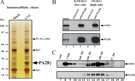
Figure 4.
The composition of coilin-containing complexes changes upon UV-C treatment. (A) Silver staining of the immunoaffinity eluates from either mock- or UV-C–treated 293 cells transiently transfected with a FLAG-coilin expression vector. Arrows indicate the protein band corresponding to FLAG-coilin and PA28γ, respectively (the latter is enriched in the complexes isolated from UV-C–treated cells). (B) Endogenous PA28γ and coilin form a high-salt–resistant complex in vivo. Nuclear extracts obtained from MCF-7 cells were immunoprecipitated with a mix of two anti-PA28γ polyclonal antibodies, as described in Materials and methods. Blotting analysis reveals the presence of both coilin and PA28γ in the immunoprecipitated material (lanes 2–3). The copurification of coilin and PA28γ is not prevented by high-salt treatment of the beads (lanes 4–5). (C) PA28γ is present in a subset of coilin-containing complexes. Nuclear extracts derived from 293 cells were fractionated by gel-filtration chromatography and subjected to Western blot analysis with anti-coilin and -PA28γ antibodies (upper and lower panels, respectively) showing that PA28γ cofractionates with a subset of coilin-containing complexes.

