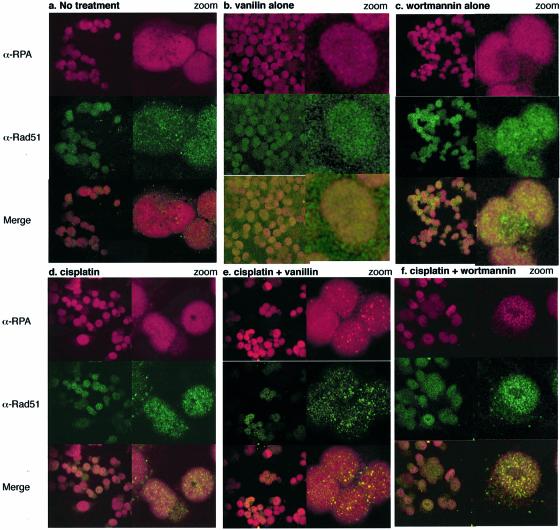
Figure 7.
Confocal microscopy. RPA/Rad51 foci formation in the nuclei of A2780 cells: (a) untreated, (b) treated with 300 mM vanillin, (c) exposed for 1 h to 25 µM wortmannin followed by 20 h incubation, (d) exposed to 5 µM cisplatin for 1 h followed by 20 h incubation, (e) exposed to cisplatin while in continuous exposure to vanillin (300 µM) and (f) exposed to cisplatin followed by a 1 h exposure to wortmannin 20 h later. RPA (red), Rad51 (green) and merge (yellow).

