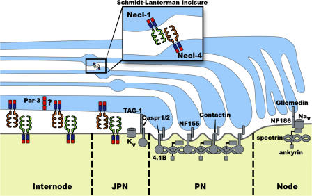
Figure 1.
Model of interactions between myelinating Schwann cells and axons showing proteins that localize to the node, paranode, juxtaparanode, and internode. Schwann cell, blue; axon, yellow. PN, paranode; JPN, juxtaparanode. Analysis of the Necl proteins provides important new insight into molecular complexes along the internode (Maurel et al., 2007; Spiegel et al., 2007), which had not been as well defined as for the other domains. The polarity protein Par-3 colocalizes with Necl-4 in the adaxonal region of the Schwann cell (Chan et al., 2006), and it is possible that these proteins interact via PDZ and PDZ-binding motifs (red). The Necl proteins can also interact with FERM domain proteins via the FERM-binding domain (blue). Necls are also present at the Schmidt-Lanterman incisures (inset). Necl-2 is not depicted but localizes to the axon–Schwann cell interface and to the incisures. The figure was adapted from Voas et al. (2007).

