Figure 1.
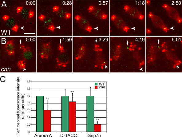
The centrioles in cnn embryos cannot maintain a proper connection to the PCM. (A and B) Still images from videos of WT and cnn syncytial embryos expressing the PCM marker GFP–D-TACC (pseudocolored red), and the centriole marker mRFP-Fzr (pseudocolored green; note that mRFP-Fzr is also concentrated in the nucleus in interphase). Time in min:s. (A) In WT embryos, the centrioles are always well centered within the PCM. (B) In cnn embryos, the centrioles are associated with PCM, but they “rocket” around within the cytoplasm and no longer maintain their proper connection to the PCM (see Video 1, available at http://www.jcb.org/cgi/content/full/jcb.200704081/DC1). (C) Quantification of the amount of PCM recruited to centrioles in WT and cnn embryos. Error bars represent SD (n = 45 for each marker and genotype). **, P < 0.001 (t test). Bar, 10 μm.

