Figure 2.
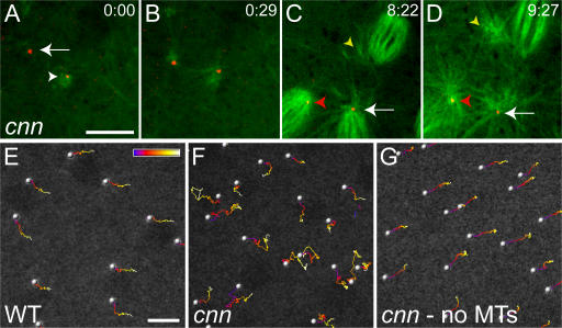
The centrioles associate with astral MTs in cnn embryos, and centriole rocketing is MT dependent. (A–D) The centrioles in a cnn embryo were visualized with mRFP-PACT (red) and the MTs with GFP–α-tubulin (green). (A) At the start of this time series (time in min:s), one centriole is associated with MTs (arrowhead), whereas another is not (arrow). The latter centriole starts to nucleate MTs from one side (B), and both centrioles exhibit “rocketing” movements as they recruit PCM and nucleate MTs asymmetrically around themselves (see Video 3, available at http://www.jcb.org/cgi/content/full/jcb.200704081/DC1). As this embryo enters mitosis (C), the centrioles continue to rocket around in the cytoplasm (Video 4). In this panel, one centriole is located at the pole of a mitotic spindle (arrow), and this pole is associated with astral MTs. Another centriole loses its connection to the spindle pole (red arrowhead) and starts to move around within the spindle; another centriole (that is not in focus, but whose position can be inferred from its associated astral MTs; yellow arrowhead) starts to migrate away from the spindle into the cytoplasm. By the end of mitosis (D), the latter centriole has completely lost contact with the spindle (yellow arrowhead); the other centriole has migrated back to the pole (red arrowhead). (E–G) Traces of centriole movement over time are shown for a WT embryo (E), a cnn embryo (F), and the same cnn embryo after colchicine injection (G). The position of each centriole over time is indicated by the color scale, which represents 150 s. See Video 5. Bars, 10 μm.

