Figure 4.
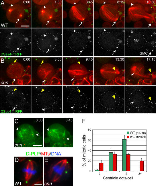
Centriole segregation is abnormal in cnn mutant larval brain cells. (A and B) Time series (min:s) of living WT and cnn mutant NBs expressing the centriole marker DSas-4–mRFP (arrows; pseudocolored green) and GFP–α-tubulin (pseudocolored red). (A) In WT cells, the centrioles are always located at the poles of the mitotic spindle. (B) In cnn NBs, the centrioles move erratically around the cell and no longer maintain their proper connection to the poles of the spindle. Note that one of the centrioles temporarily moves out of the focal plane (13:30) but reappears by the end of mitosis. (C) Another cnn NB expressing the same markers as in A and B (colors are inverted). Both centrioles are able to nucleate astral MTs but fail to maintain their connection to them. (D and E) The distribution of MTs (red), centrioles (D-PLP; green), and DNA (blue) in fixed WT and cnn mutant cells. In cnn mutant cells (E), the centrioles are more randomly distributed in the cell and are sometimes of unequal size (which may reflect the clustering of some centrioles). (F) Bar chart showing the number of centrioles present in WT and cnn mutant mitotic brain cells. Error bars represent SD. Bars, 5 μm.

