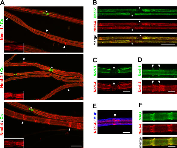
Figure 3.
The Necl proteins are localized in the internode and Schmidt-Lanterman incisures. (A) Teased sciatic nerves from adult mice were stained with antibodies to Necl-1, -2, or -4 (red) and Caspr (green), a marker of the paranodes. Antibodies to all three Necl proteins stain the internode and Schmidt-Lanterman incisures (arrowheads). Nodes, which are indicated by asterisks, are shown magnified in the insets without Caspr staining to demonstrate that Necl expression is largely excluded from the paranodes. (B) Teased sciatic nerves were double stained for Necl-1 (green) and -4 (red); the merged image is shown below. Two nodes and their flanking paranodes located in the center of the field are indicated with asterisks and are unlabeled. (C) A node of Ranvier stained for Necl-1 (green) and -4 (red). Both Necl-1 and -4 are largely excluded from the paranodes and node, which is indicated with asterisks. (D) An internodal segment of a myelinated nerve shows two incisures (arrowheads) that are stained with antibodies to Necl-1 (green) and -4 (red). In each case, Necl-1 expression is restricted to the outer portion of these clefts, whereas Necl-4 stains the entire cleft. (E) A segment of a myelinated nerve stained for Necl-4 (red) and MBP (blue) is shown. The arrowhead indicates a Schmidt-Lanterman incisure. (F) Necl-4 (red) colocalizes with MAG (green) along the glial internode and in the clefts as shown. Bars (A and B), 25 μm; (C–F) 10 μm.

