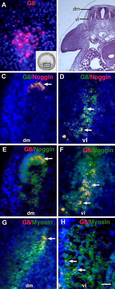
Figure 1.
Expression of Noggin and myosin in MyoDpos cells originating in the epiblast. MyoDpos cells labeled with the G8 mAb were present in the posterior region of the stage 2 epiblast (red cells in A). 4–5 d after labeling with G8, stage 25 embryos were examined for expression of Noggin and sarcomeric myosin. Regions indicated on the right side of the embryo in the hematoxylin and eosin–stained section are shown at higher magnification in fluorescence photomicrographs of merged images of G8 mAb (labeled with Alexa Fluor 488 [green in C and D] and rhodamine [red in E–H]) and either Cy3/red-labeled dendrimers to Noggin mRNA (C and D) or Alexa Fluor 488/green–labeled antibodies to Noggin (E and F) or myosin (G and H). Nuclei were stained with Hoechst dye (blue). Double-labeled cells (overlay of red and green) appear yellow (arrows). (C–F) G8pos/ Nogginpos cells were observed in the dorsomedial (dm) and ventrolateral (vl) dermomyotome and myotome. (G and H) G8pos/myosinpos cells were present in the myotome. Bar: (A and C–H) 9 μm; (B) 135 μm.

