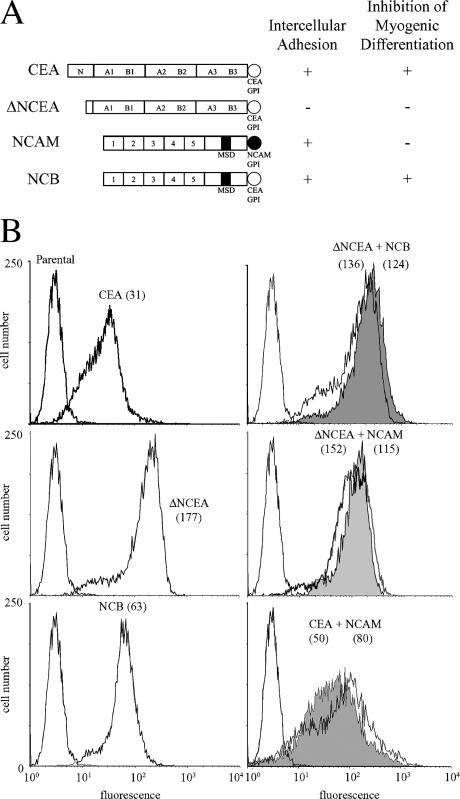
Figure 1.
Surface expression of CEA and NCAM proteins on L6 myoblasts. (A) Schematic representation of proteins used in this study, with their ability to mediate intercellular adhesion and to inhibit myogenic differentiation shown (as + or −). (B) FACS profiles, after staining with mAbs D14 (anti-CEA) or 123C3 (anti-NCAM) and FITC-conjugated secondary antibody, demonstrate cell surface levels of indicated proteins with fluorescence means in parentheses. In coexpressing cells, D14 staining is shown by shaded profiles.

