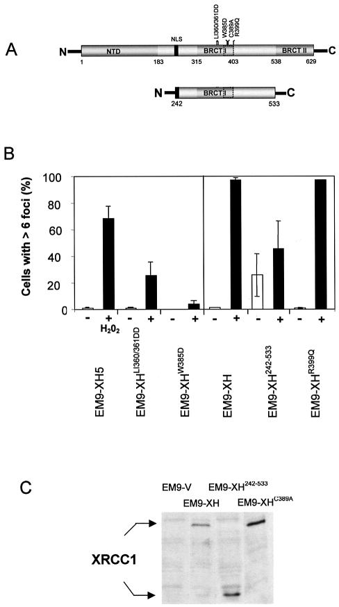
Figure 3.
The BRCT I domain is required for appearance of XRCC1 nuclear foci after oxidative DNA damage. (A) Schematic of full-length XRCC1 (top) and XRCC1242–533 (bottom) depicting the proposed PAR- binding motif (dotted box) located within BRCT I domain. The position of the nuclear localization signal (NLS) and of the mutations employed in this study are shown. (B) Mutation of the BRCT I domain inhibits assembly of XRCC1 nuclear foci after H2O2 treatment. The indicated cell lines were mock-treated (‘–’) or treated (‘+’) with H2O2 for 20 min, incubated in drug-free medium for 10 min, and then processed for anti-XRCC1 immunofluorescence as described for Figure 1. 100 cells from each cell line were scored for the presence or absence of six or more foci. Each data point represents the mean of two independent experiments and error bars reflect the data range. EM9-XH5 is a single transfectant clone and is the wild-type control for EM9-XHLI360/361DD and EM9-XHW385D, with which it expresses similar levels of recombinant human XRCC1 (25). EM9-XH is a pooled population of more than 50 transfectants and is the control cell line for EM9-XH242–533 and EM9-XHR399Q (25). (C) XRCC1 protein levels in EM9-V, EM9-XH, EM9-XH242–533 and EM9-XHC389A cells. Protein extracts from the indicated cell lines were fractionated by SDS–PAGE, transferred to nitrocellulose and immunoblotted with anti-XRCC1 polyclonal antibody.

