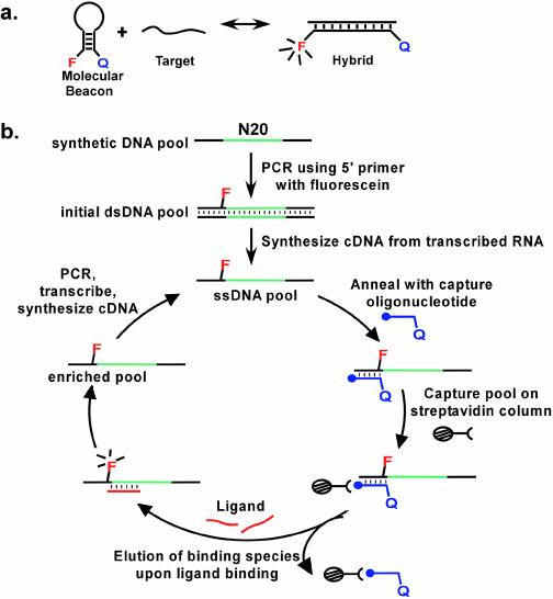
Figure 1.
Selection scheme for molecular beacons. (a) Conformational changes in designed molecular beacons. F represents an embedded fluorophore, Q a quencher. (b) In vitro selection of molecular beacons. The closed circle at the termini of the capture oligonucleotide represents biotin. The selection protocol is described in greater detail in Materials and Methods, and Results and Discussion.

