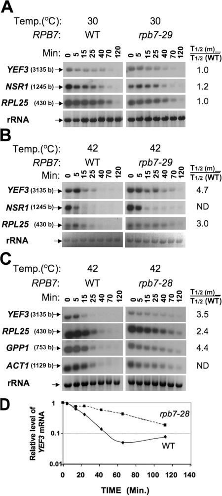
Figure 2.
PBF and non-PBF mRNAs decay abnormally slowly at the nonpermissive temperature in rpb7 ts mutants. (A) At the permissive temperature, rpb7-29 and WT exhibit similar mRNA decay kinetics. The indicated strains were proliferated at 30°C until mid-log phase. 1, 10, phenanthroline was added to block transcription (Grigull et al., 2004; Lotan et al., 2005). Decay kinetics was determined by monitoring mRNA levels at the indicated time points post-transcription block, using Northern analysis with the indicated probes. rRNA serves to demonstrate equal loading. Half-lives (designated T1/2) were determined as described previously (Lotan et al., 2005) and the ratios between T1/2 of the mutant and that of the wild-type [T1/2 (m)/T1/2 (WT)] are indicated on the right. Lengths of the open reading frames are indicated in parentheses next to the gene name on the left. (B) At the nonpermissive temperature, rpb7-29 cells exhibit defective mRNA decay. Cells were challenged simultaneously with 1, 10, phenanthroline and shifted to 42°C and mRNA decay was determined as in A. (C) rpb7-28 cells are defective in mRNA decay. Cells were proliferated at 30°C till mid-log phase and then shifted abruptly to 42°C (no drug was added). The high temperature led both to inactivation of Rpb7-28p ts and to transcription arrest (Lotan et al., 2005). mRNA decay was determined as in A. (D) Decay kinetics of YEF3 mRNA in WT and rpb7-28 cells was determined as described previously (Lotan et al., 2005).

